The time has come to talk about an innovative and interesting topic, namely dermatoscopy.
Tell us in a few words about this method.
This is one of my main and most favourite fields, namely dermatoscopy of skin growths.
What is the dermatoscope and the dermatoscopy procedure itself?
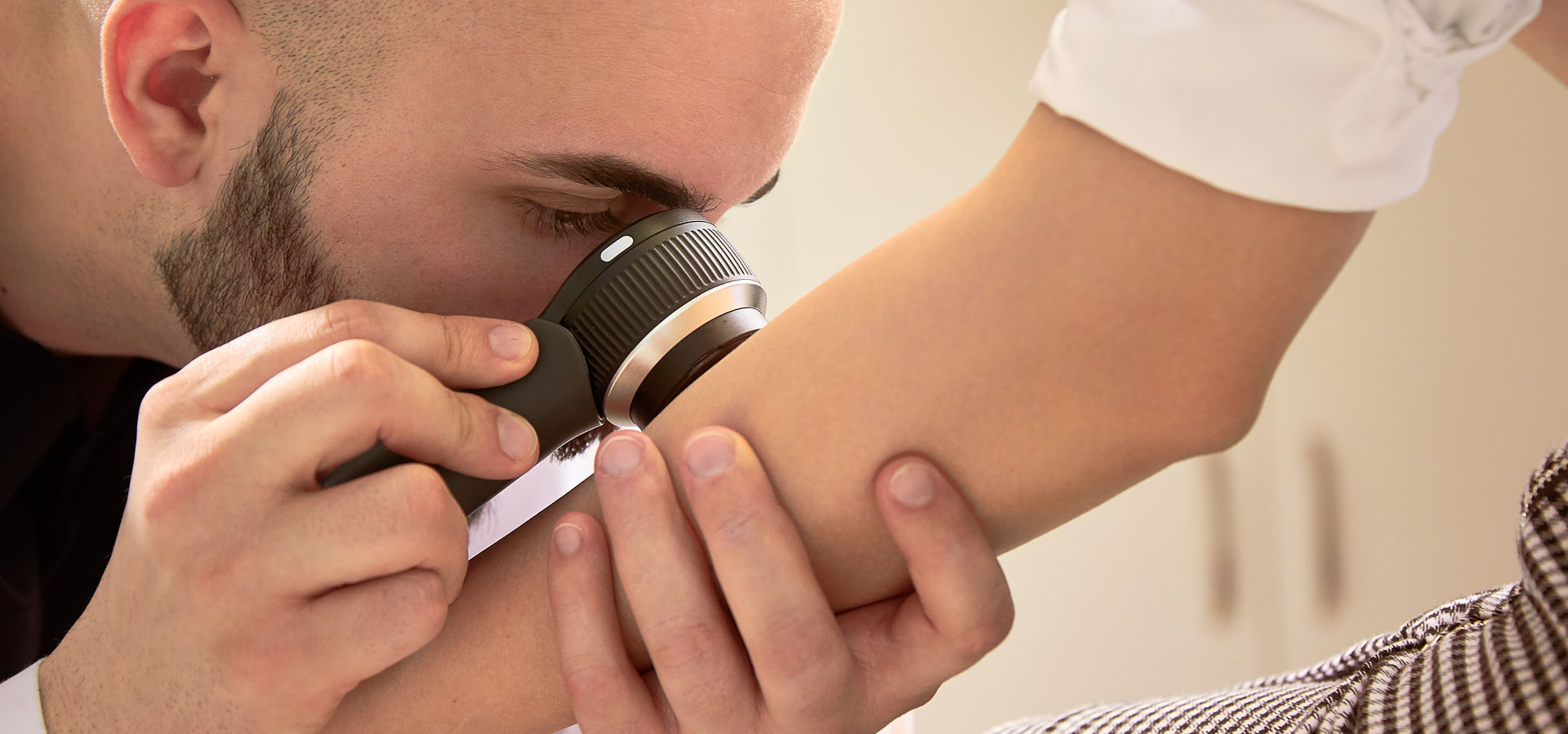
Dermatoscopy is a non-invasive method that very easily and affordably allows us physicians to observe in vivo the structures of the epidermis, dermo-epidermal border and papillary dermis. These structures are directly related to the histological characteristics of the corresponding lesion.
The dermatoscope itself consists of a transparent plate, a light source (polarized or nonpolarized), and a magnifying glass. Some of the newer models allow digital recording and image capture. The magnification of the observed image through the dermoscope varies between 10 and 100 times.
What are the advantages of this method?
The advantages are many, but I will mention the most important ones. In most cases, the equipment is very portable and convenient to use. With a qualified specialist, the procedure is highly specific, and in a significant number of cases, unnecessary dermatohistopathological examination can be avoided. The technique is rapid and a large number of skin lesions can be examined in a short period of time. It allows digital recording of the lesions and their follow-up over time.
What are the most common growths you diagnose with this method?
The most important differentiation in my opinion is that between a benign melanocytic nevus and a malignant melanoma. In some of the lesions, the line is very thin and with this method we can catch very early neoplastic transformation and save lives. Another type of lesion that we can easily and quickly diagnose is basal cell carcinoma. The smallest such lesion I have diagnosed, excised and dermatohistopathologically verified is a 2mm basal cell carcinoma in the suprapubic zone. We also see benign lesions such as seborrheic keratoses, fibromas, cysts, dermatofibromas and some rarer ones such as syringomas, trichoepitheliomas, hydrocystomas, etc. on a daily basis.
Can you tell us about a more interesting case from your practice where dermatoscopy was helpful?
I recently diagnosed a young man with phthiriasis caused by Phtirus pubis (pubic louse) that had been unnoticed by another colleague. This type of infectious dermatosis can easily be missed by not using the dermatoscopic method.
What is the age of the youngest patient diagnosed with skin cancer using the dermatoscopic method?
The youngest patient I have diagnosed with melanoma is 18 years of age. A young lady who had no idea the neoplasia existed. Another recent interesting case from my practice was the dermatoscopic differentiation of Spitz nevus from malignant melanoma in a 5 year old girl.
How long is the mole examination?
This of course depends on the number and type of growths. There are people who suffer from what is called Nevus Dysplasticus Syndrome, in which a large proportion of the nevi are atypical. In this type of patient, we have to be extremely careful, as it can be easy to miss an existing malignant melanoma. The standard duration of the examination is about 20 minutes.
How often do you advise people to check their moles?
I divide my patients into four groups. Those with low risk. I examine them once a year; Those with medium risk. I check them twice a year. Those with high-risk. They should be examined every 3 months. Patients operated for malignant melanoma. In most cases in the first year, these patients visit my office monthly.
Is dermatoscopy used for skin pathologies other than skin growths?
In recent years, great minds in the field have been pushing a very new concept, namely dermatoscopy of skin diseases. Some of these can only be diagnosed timely and accurately by dermatoscopy. Some of them are psoriasis, lupus, lichen planus, dermatitis, urticaria, alopecia, dermatomyositis, scleroderma and others.
Is dermatoscopy a sophisticated method in your opinion?
Some of my colleagues deny dermatoscopy. For me, it is an integral part of my practice. I have at least 4-5 patients daily for dermatoscopic examinations. The deeper I get into the subject, the more challenging, yet interesting and useful, I find it.
Give one last piece of advice to patients!
Prophylaxis, prevention, self-monitoring and if necessary consultation with a specialist! In case of even the slightest suspicion of a strange new skin lesion or a change in an old one, immediately seek a specialist profiled in dermatoscopy.
We use information collected through cookies and similar technologies to improve your experience on our site, analyse how you use it and for marketing purposes.
We and our partners use information collected through cookies and similar technologies to improve your experience on our site, analyse how you use it and for marketing purposes. Because we respect your right to privacy, you can choose not to allow some types of cookies. However, blocking some types of cookies may impact your experience of the site and the services we are able to offer. In some cases, data obtained from cookies is shared with third parties for analytics or marketing reasons. You can exercise your right to opt-out of that sharing at any time by disabling cookies.
These cookies and scripts are necessary for the website to function and cannot be switched off. They are usually only set in response to actions made by you which amount to a request for services, suchas setting your privacy preferences, logging in or filling in forms. You can set your browser to block oralert you about these cookies, but some parts of the site will not then work. These cookies do not store any personally identifiable information.
Analytics
These cookies and scripts allow us to count visits and traffic sources, so we can measure and improve the performance of our site. They help us know which pages are the most and least popular and see how visitors move around the site. All information these cookies collect is aggregated and therefore anonymous. If you do not allow these cookies and scripts, we will not know when you have visited our site.
Marketing
These cookies and scripts may be set through our site by our advertising partners. They may be used by those companies to build a profile of your interests and show you relevant adverts on other sites. They do not store directly personal information, but are based on uniquely identifying your browser and internet device. If you do not allow these cookies and scripts, you will experience less targeted advertising.